

Paratuberkulose-Sanierung durch eine Kombination von ‚Test and cull‘, Impfung und mutterloser Aufzucht – Beobachtungen in drei deutschen Milchziegenherden
Berliner und Münchener Tierärztliche Wochenschrift 136, 1–11
DOI: 10.2376/1439-0299-2022-24
© Schlütersche Fachmedien GmbH. 2023
Eingereicht: 19. November 2022
Akzeptiert: 5. Juni 2023
Publiziert: 07/2023
Summary
Three German dairy goat herds infected with Mycobacterium avium subsp. paratuberculosis (MAP) were accompanied during the trial of paratuberculosis sanitation over three years. The development of the number of goats excreting MAP was documented. On all three farms, all goats older than 1.5 years were examined annually for MAP excretion by faecal culture, and all MAP-shedders should be culled. On farms 1 and 2, initially additional blood samples were analysed by ELISA and the reactors were also culled. Due to the paratuberculosis vaccination that had already been carried out on farm 3, this was omitted here. After the first herd testing, all goats from farms 1 and 2 and the offspring from farm 3 were vaccinated with Gudair®. In the following years, the offspring of all farms were vaccinated annually. All farms were advised to separate the kids from their mothers immediately after birth and raise them motherless.
At the initial examination of the herds, the within-herd faecal culture prevalence (FCP) for MAP in the three farms was 19.1%, 26.4% and 9.1%, respectively. On farms 1 and 2, the FCP decreased significantly to 2.0% and 8.9%, respectively. On farm 3, FCP was reduced to 6.7%, but the difference from baseline was not significant.
The procurement of sufficient quantities of MAP-unsuspicious colostrum and milk for motherless rearing, but also the identification and culling of MAP-positive animals as well as the rapid separation of the kids from their mothers after birth and the strict segregation of the different age groups proved to be problematic. Consistency of the farm managers in implementing the measures and good herd management seem to influence the success of sanitation. The observations in the three herds show, that the combination of the control measures test and cull, vaccination and motherless kid rearing can considerably reduce the prevalence of MAP shedding goats in a relatively short period of time, but complete sanitation could not be achieved.
Zusammenfassung
Drei verschiedene deutsche Milchziegenherden, die mit Mycobacterium avium subsp. paratuberculosis (MAP) infiziert waren, wurden bei dem Versuch der Paratuberkulose-Sanierung über drei Jahre begleitet. In allen Betrieben wurden jährlich alle Ziegen älter als 1,5 Jahre mittels kultureller Untersuchung von Kotproben auf die Ausscheidung von MAP untersucht und alle Ausscheider sollten gemerzt werden. In den Betrieben 1 und 2 wurden initial zusätzlich Blutproben serologisch mittels ELISA untersucht und die Reagenten ebenfalls gemerzt. Aufgrund bereits jahrelang durchgeführter Paratuberkulose-Impfung in Betrieb 3 entfiel dies hier. Nach der ersten Herdenuntersuchung wurden alle Ziegen von Betrieb 1 und Betrieb 2 sowie die Nachzucht von Betrieb 3 mit Gudair® geimpft. Anschließend wurde in allen Betrieben jährlich jeweils die Nachzucht geimpft. Alle Betriebe sollten die Kitze unmittelbar nach der Geburt von den Müttern trennen und mutterlos aufziehen.
Bei der initialen Untersuchung der Herden lag die Innerherdenprävalenz der kulturellen Kotuntersuchung in den drei Betrieben bei 19,1, 26,4 bzw. 9,1 %. In Betrieb 1 und Betrieb 2 sank die Innerherdenprävalenz signifikant auf 2,0 % bzw. auf 8,9 %. In Betrieb 3 konnte die MAP-Prävalenz auf 6,7 % reduziert werden, der Unterschied zur Ausgangssituation war allerdings nicht signifikant.
Als problematisch erwiesen sich die Beschaffung ausreichender Mengen an MAP-unverdächtigem Kolostrum und Milch für die mutterlose Aufzucht, aber auch die Identifizierung und Keulung MAP-positiver Tiere, das zügige Trennen der Kitze von den Müttern sowie die Einhaltung einer strikten räumlichen Trennung der verschiedenen Altersgruppen. Konsequenz der Betriebsleiter bei der Umsetzung der Maßnahmen und ein gutes Herdenmanagement scheinen den Erfolg der Bekämpfung entscheidend zu beeinflussen. Die Beobachtungen in den drei Herden zeigen, dass durch die Kombination der Bekämpfungsmaßnahmen ‚Test and Cull‘, Impfung und mutterloser Lämmeraufzucht die MAP-Prävalenz in relativ kurzer Zeit deutlich reduziert werden kann, eine vollständige Sanierung wurde allerdings nicht erreicht.
Introduction
This case report describes the trials to reduce the prevalence of infections with Mycobacterium avium subsp. paratuberculosis (MAP) in three different German dairy goat herds.
Paratuberculosis or Johne’s disease (JD) occurs worldwide in ruminants and is widespread in German dairy goat herds (Stau et al. 2012). The disease induces not only relevant losses in milk yield in the affected herds, but also represents a clinical and animal welfare problem. The chronic granulomatous intestinal disease is caused by Mycobacterium avium subsp. paratuberculosis. Goats have a lower natural resistance to MAP than sheep and cattle and are therefore more susceptible to infection (Stewart et al. 2007). It is estimated that up to 71% of goat herds in Germany are infected with MAP (Stau et al. 2012). In that prevalence study, the goat herds were mainly kept for milk production, but goats from mixed herds with sheep for landscape preservation were also included (Stau et al. 2012).
The pathogen is excreted by infected animals mainly in the faeces, but excretion via colostrum and milk have also been described (Windsor 2015). Infection occurs through oral ingestion mainly by faecal contaminated feed, pasture and drinking water (Sweeney 2011). In utero infection in a heavily infected goat is also reported (Alinovi et al. 2009). Young animals are particularly susceptible; later, a certain age resistance develops, so that higher infection doses are necessary for infection (Sweeney 1994, Clarke 1997, Fecteau et al. 2010, Sweeney 2011).
Top Job:
The incubation period lasts several months to years, depending on the dose of infection and the age of the animal. The clinical signs are progressive weight loss with initially undisturbed feed intake. Diarrhoea is not typical in goats (Djønne 2010). In the final stage, affected animals dehydrate, become anaemic and develop dysproteinaemia with hypalbuminaemia and possibly hypergammaglobulinaemia (Schroeder et al. 2001, Bonelli et al. 2017), followed by submandibular oedema and increasing weakness. Eventually, the disease is fatal. The more advanced the disease is, the more pathogens are excreted (Djønne 2010).
In infected herds, economic losses occur due to higher susceptibility to diseases, poor feed conversion, reduced milk yield and fertility, and higher mortality rates (Kostoulas et al. 2006a, Sardaro et al. 2017).
Due to the non-specific clinical signs, further diagnostics are essential. The cultural examination of faeces is considered as gold standard. With a cross-species specificity of 0.97–1.0 (Nielsen and Toft 2008), reliable identification of shedders is possible. However, the culture is not very sensitive and therefore many shedders may remain undetected. Data on the sensitivity of faecal culture in cattle vary from 0.23 to 0.74 depending on the stage of infection (Nielsen and Toft 2008). The sensitivity of faecal culture in goats varies between 0.08, 0.27 and 0.71 depending on the population studied and its age structure (Kostoulas et al. 2006b, Mercier et al. 2009, Hemati et al. 2022).
Besides faecal culture, serological blood testing by ELISA is frequently used, as these tests have a high throughput and are relatively inexpensive. The sensitivity of the different commercial ELISAs in goats varies between 0.015 (Mercier et al. 2009) and 1.0 (Molina Caballero et al. 1993), depending on the population examined and the ELISA used. Clinically ill animals are better detected than subclinical ones. The specificity varies between 0.79 (Dimareli-Malli et al. 2004) and 1.0 (Whittington et al. 2003b) in goats (Nielsen and Toft 2008). To increase the overall sensitivity, different test procedures should be combined and tests repeated regularly.
According to Rerkyusuke and Ganter (2015) the sensitivity and specificity of the ELISA used here (Cattletype® MAP Ab [Indical Bioscience GmbH, Leipzig, Germany]) for goats is 84% and 100% respectively. For cattle, sensitivity and specifity of the same ELISA were 53.6% and 99.3%, respectively (Koehler et al. 2008).
In the early incubation phase, in vivo diagnostics are unreasonable, as these goats are not yet shedding MAP. Furthermore, even in the subclinical stage of disease, diagnosis is still very uncertain (Rerkyusuke et al. 2019). No precise statement can be made after which period of time shedding takes place. In this study, the goats were only tested from the age of 18 months on.
To control JD, it is necessary to interrupt transmission routes and reduce the infection pressure.
According to Windsor (2015), the three main approaches to control paratuberculosis in small ruminants are:
- management measures to reduce transmission,
- test and cull to eliminate sources of infection, and
- vaccination of replacement animals to increase their resistance to infection.
Identifying and culling infected animals is an effective measure to reduce infection pressure, as MAP-excreting animals excrete up to 108 organisms per gramme faeces (Reddacliff et al. 2006). The higher the infection pressure, the greater the risk that older animals will also become infected. The risk that infected animals remain undetected in the herd is present due to the limitation of the tests. However, as the testing procedures become more accurate for the progressive stages of the infection (Nielsen and Toft 2008), the animals with the highest potential for transmission are most likely to be detected (Robbe-Austerman 2011).
Test and cull or reagent eradication is a component of most international JD control programmes, but this alone is insufficient to control infection. Without further measures to interrupt the infection cycle, infection transmission will continue (Geraghty et al. 2014, Strain 2018).
Reduction of the exposure of young animals to faeces of pathogen-excreting adults or MAP-contaminated feed is also the focus of many control programmes (Strain 2018). Separate motherless rearing of the particularly susceptible young animals in a clean environment is therefore an option to interrupt the infection chain. Kids should be raised on MAP-free colostrum and milk (Djønne 2010, Windsor 2015).
Another possibility to reduce the infection pressure is vaccination. Currently, only the inactivated vaccine Gudair® (CZ Veterinaria [now CZ Vaccines], O Porriño [Pontevedra], Spain) is licensed in EU-member states. The vaccine contains the inactivated MAP strain 316F and mineral oil as adjuvant. A single vaccination is sufficient.
Vaccination induces both a cellular and humoral immune response and reduces shedding of MAP bacteria by 90% (Reddacliff et al. 2006). The onset of the disease is delayed and thus the incidence of clinical cases is decreased. Mortality from JD was reduced by 90% through vaccination. Nevertheless, there remains a risk that vaccinated animals will become ill and transmit the disease (Corpa et al. 2000, Reddacliff et al. 2006).
As vaccination only reduces MAP contamination but does not allow complete eradication, routine vaccination of the offspring is necessary to maintain control over the incidence of infection (Dhand et al. 2016).
Due to its good cost-benefit ratio, vaccination is an effective way to decrease contamination with MAP, to reduce clinical disease, and to maintain productivity in dairy goat herds. Vaccination can be used to keep the disease at a low level (Juste and Perez 2011).
The following case study focuses on three different German dairy goat herds that experienced JD as a herd problem and how they tried to control it. The implementation of the preventive measures test and cull, vaccination and motherless kid rearing as well as the reduction of the within-herd prevalence of MAP shedders as determined by faecal culture are described. Moreover, the successes achieved in a three-year period, but also the problems in implementing the aforementioned measures are discussed.
Case Description
Farms
The three observed dairy goat farms are located in North-Rhine-Westphalia (Germany) and were accompanied between 2017 and 2020. They are no experimental herds, but normal farming operations.
The initial situation in the herds before measures against paratuberculosis were taken is shown in Table 1.
As evidence of MAP was available in all three farms, a plan of measures was drafted with the goal to reduce and if possible, eliminate MAP-infections. Three sanitation measures had to be implemented on all farms.
In order to reduce the number of MAP-infected and MAP-excreting goats, the animals had to be regularly examined and reactors had to be culled. For this purpose, on farms 1 and 2, blood and faecal samples were initially collected in 2017 from all goats older than 1.5 years. On farm 3, only faecal samples were collected, as due to prior vaccination with Gudair® (Table 1), serological examination was omitted.
Serum monovettes (Sarstedt AG & Co. KG, Nuembrecht, Germany) were used for blood sampling from the Vena cava cranialis or the jugular vein. After centrifugation (2000g, 15 minutes, centrifuge Hermle Z326, HERMLE Labortechnik GmbH, Wehingen, Germany), the serum was analysed for MAP antibodies using Cattletype® MAP Ab ELISA (Indical Bioscience GmbH, Leipzig, Germany) in accordance with the manufacturer’s instructions in the laboratory of the Clinic for Swine and Small Ruminants of the University of Veterinary Medicine Hannover, Foundation, Hannover, Germany (Clinic). In accordance with the manufacturer’s instruction, the cut-off for cattle, sheep and goats is at an S/P ratio (ratio of optical density of the sample to mean optical density of the positive control) of 0.4. To increase sensitivity, the cut-off of the S/P ratio was reduced from 0.4 to 0.25 (Rerkyusuke and Ganter 2014). A loss of specificity was accepted here, as it is crucial for rapid sanitation that as few goats as possible with a false negative test result remain in the herd and are recognised early as potential MAP-shedders.
The faecal samples were taken rectally with disposable gloves and examined for the presence of MAP at the Chemical and Veterinary Investigation Office Rhein-Ruhr-Wupper (CVUA-RRW, Krefeld, Germany) by means of cultural examination in accordance with the standard procedure of the German official collection of methods of the Friedrich-Loeffler-Institut (Federal Research Institute for Animal Health) (Anonymous 2020). Incubation was carried out for up to nine months.
Since the results of the ELISA were available more quickly, all goats with a reactivity above the cut-off value of S/P>0.25 were culled. Animals that were negative in the ELISA, but subsequently found to excrete MAP in their faeces were also removed from the farm. On farm 3, the culling decision depended solely on faecal culture results. The bacterial growth was monitored regularly during the nine-month incubation, and as not all cultures grew at the same time, the results were available at different times, so that culling took place gradually.
Subsequently, annually until 2020, all goats from the age of 1.5 years upwards were tested for MAP excretion by individual faecal culture on all three farms and positive goats were culled. If an animal was pregnant when the positive result arrived, the animal was culled immediately after kidding.
It was communicated with the farmers that the purchase of new animals should be avoided if possible, new breeding bucks should be at least 1.5 years old and serologically tested for paratuberculosis immediately after arrival on the farm. If the animals had already been vaccinated, they were tested for MAP in faecal samples. Until a result was available, the bucks were kept in quarantine.
In order to reduce pathogen excretion in undetected infected goats, all goats on farms 1 and 2 were vaccinated subcutaneously on the lateral chest wall with 1 mL of Gudair® after the initial examination. Since the herd on farm 3 had already been vaccinated since 2010, only the offspring was vaccinated in this case. As the vaccine was not licensed in Germany, an exemption permit for the herds was granted by the state of North Rhine-Westphalia, Germany, in accordance with § 11 para. 6 no. 2c of the German Animal Health Act. According to the manufacturer a singular injection of Gudair® is sufficient to induce protective cellular and humoral immunity against MAP. Therefore, in the following years, only the offspring aged 3–8 months was vaccinated on all three farms.
Since vaccination leads to seroconversion in non-MAP-infected animals (Stau and Ganter 2012), further identification of infected animals by ELISA was no longer useful.
To interrupt the infection chain from excreting does to the susceptible young animals, the kids were reared motherless in a separate building on all three farms. It was planned to separate the kids from their dams immediately after birth and feed them with MAP negative cow colostrum. But due to the high work load and lack of employees, this plan was not always fulfilled to 100%. Especially on farm 2 and to a latter extend on farm 3 immediate separation after lambing was carried out more or less consequently. The colostrum used, came from a dairy cattle herd that had been tested negative for JD over several years. Beginning with day two, the kids were fed with whole milk powder or milk replacer until weaning. This procedure was continued throughout the entire period.
Separating goat kids from their dams took place on farms 1 and 3 for approx. 12 months and the young animals were only integrated into the herd of adult goats at the first kidding. On farm 2, the separation was only possible for approx. 6–8 months for operational reasons. Afterwards, the young goats were reared in an extra group next to the adult goats in the same barn.
Results
Table 2 shows the detailed results of each testing in the three herds. The apparent MAP within-herd prevalence is described in relation to the number of evaluable samples.
Farm 1 had an initial prevalence of 23.6% when ELISA and faecal culture were combined and farm 2 of 40.9%. The results of the two tests overlapped only in parts (Figure 1). For better comparability with farm 3, the focus was placed on the within-herd faecal culture prevalence (FCP).
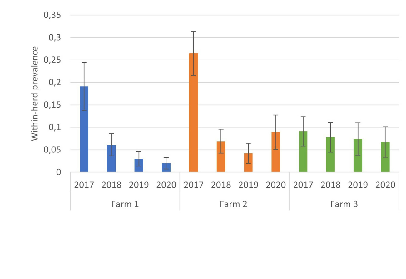
This was 19.1% (95%CI = [13.8–24.4%]) for farm 1, 26.4% [21.5–31.3%] for farm 2 and 9.1% [5.8–12.4%] for farm 3 in the first testing. In the following years, the FCP on farm 1 decreased to 6% [3.6–8.4%] after one year, then to 3% [1.3–4.7%] and in the last year MAP excretion could only be detected in 2% [0.7–3.3%] of the goats.
On farm 2, the FCP decreased to 6.9% [4.2–9.6%] after one year, then to 4.2% [2.0–6.4%] and increased again to 8.9% [5.1–12.7%] in the last year. On farm 3, the prevalence dropped over 7.8% [4.4–11.0%] and 7.4% [3.8–11.0%] to 6.7% [3.3–10.1%] (Figure 2).
A statistical analysis of the prevalence data was done by Pearson’s Chi-squared test. The difference between the FCP in the initial situation and at the end of the study was significant on farms 1 (p<0.001) and 2 (p<0.001) but not on farm 3 (p=0.3).
In all herds not all samples from the tested goats were evaluable. Either an insufficient amount of faeces could be obtained or the cultivation was overgrown by other agents and thus not analysable. The FCP was calculated on the basis of the evaluable samples and not on the number of tested goats.
It proved difficult to identify and remove all the positive goats in time. On farm 1, after the first sampling not all positive goats were removed from the farm which, however, only became apparent during the sampling in the following year. The same happened after the third testing where one goat was not removed. On farm 2, five positive goats from the first examination were still in the herd at the time of the second testing, and at the third testing again, six positive goats from the previous sampling were still in the herd. On farm 3, at the time of the final testing, two goats that had already shown a positive result in the previous year were still in the herd.
The positive goats that were not removed were in all cases tested in the following year. Not in all cases did the culture give a positive test result again, but all these goats were still considered to be infected and were then culled.
Herd 1 had continuously grown due to its own offspring. The implementation of motherless rearing was very consistent and the general farm hygiene was good (Table 3).
After 40% of the goats on farm 2 had been culled in 2017, the farmer decided to purchase additional goats for operational reasons. In spring 2018, a total of 148 approximately nine-month-old goats were purchased from a Dutch breeding farm, where they should have already been vaccinated with Gudair® as young kids. To ensure full vaccination coverage, these young goats were vaccinated a second time with Gudair® when they arrived at the farm. Even though these goats were not yet 1.5 years old at the time of the second herd testing (2018), they were still sampled to identify a possible MAP introduction at an early stage. Nonetheless, these animals all showed a negative faecal culture result at this testing. At the testing in 2019, one of the purchased goats and in 2020 four of the purchased animals were tested MAP positive.
Motherless rearing was often practiced only up until day two or three in the male kids. However, the male kids left the farm at around day 14 of life. The female kids were separated from their mothers within the first day of life. It was not always possible to avoid suckling of colostrum from the kids at their dam.
In the later rearing period, a clear segregation within the individual barns could not always be guaranteed as some goats were roaming free. This resulted in contact between the young goats and the adult animals or their excreta.
On farm 3, the male kids also stayed with their mothers for 1–3 days but were housed in a separate pen from the female kids in the rearing barn and usually left the farm at 14–28 days of age. Also on this farm, the female kids were not always separated from their mothers immediately after birth, but the separation usually took place within the first day of life.
In 2018, during the testing of the cattle farm from which the colostrum and also the whole milk, with which the kids on this farm were reared, it turned out that five of the 21 cows were serologically MAP-positive. However, MAP excretion with the faeces could not be detected. In the following years, the colostrum for the kids was obtained from another cattle farm, which was regularly tested serologically MAP-negative and from which the other two farms also obtained their colostrum. Further rearing was done with milk replacer.
The differences in management and the problems occurring when implementing the measures are shown in Table 3.


Discussion
The aim of this study was to find out which effects the measures test and cull, motherless rearing and vaccination with Gudair® have on the within-herd prevalence in the three naturally infected dairy goat herds in a three-year period.
Due to the poor comparability of the different test methods at the beginning, the case study focused on the faecal culture prevalences. Nevertheless, it was important to perform the ELISA on the farms if possible, as more animals could be identified as positive and thus as a source of infection.
The FCP could be reduced on all farms, but complete sanitation was not achieved. A significant difference between the FCP before the introduction of the measures (first testing) and after the implementation of the measures over 3 years (last testing) existed for farm 1 and 2, but not for farm 3. The higher the initial prevalence, the faster a significant reduction seems to be achieved. However, a further decrease took place gradually.
Faecal culture prevalence on farm 3 has increased before the study period from 6% in 2013 to 9,1% in 2017 despite the fact, that the herd was vaccinated since 2010. The reasons for this can only be speculated. Prior to the study, the kids received colostrum directly from their dams and were then raised on whole milk from a cattle farm whose MAP status was unknown. This might be a possible source of infection. Due to the low sensitivity of faecal culture, it is also possible that some infected goats were simply not detected in the 2013 testing or were not shedding MAP at that time. Another reason might be, that the time of the examination plays a role. In this study, the goats were tested after the end of the lambing season. After this stressful time the excretion rate might be higher than at any other time of the year. Nevertheless, these points are only assumptions and not a well-founded explanation for an increase in prevalence despite vaccination.
In this investigation farm 1 showed the greatest reduction in FCP with 2.0% at the end of the study. This farm had good farm hygiene and was particularly consistent with implementing motherless rearing. Except for three does, the positive-tested goats were culled immediately after the test results were received. However, it must be considered that the herd has grown due to its own offspring and therefore there are more younger goats in the herd. These have a lower probability of excreting MAP.
On farm 2, there was an increase in prevalence again from 2019 to 2020. On farm 3, FCP was only slightly reduced.
Factors that may have led to a worse result in farms 2 and 3 compared to farm 1 are:
- access to pasture, as due to the high tenacity of MAP, grazing is very likely to pose a risk for new infections (Whittington et al. 2003a, Fecteau et al. 2010)
- single MAP excretors remaining in the herd especially on farm 2
- kids staying up to 3 days with the dams
- on farm 2, numerous free-roaming goats between the groups and on the feeding table
- on farm 3, feeding the kids with milk from serologically MAP-positive cows during the first year of sanitation.
The compliance of the farm managers is an essential factor for successful sanitation. Inconsistency in motherless rearing and in immediate culling of MAP shedders led to a delay in the sanitation process on farm 2. Also on farm 2, a carry-over of MAP between the age groups cannot be ruled out, as a safe segregation of young goats and adult goats was not always guaranteed and contact occurred here due to goats escaping from their pen and even barn. In addition, a notably large number of cultures on this farm could not be evaluated due to overgrowth with other agents. The reason for this remained unclear. Feed quality may have played a relevant role. As a result, a not inconsiderable proportion of MAP-shedders may have remained undetected in the herd and led to a further delay in sanitation. As the prevalence was determined in relation to the evaluable faecal samples, there may be a falsification here, as the herd actually consisted of more animals.
General farm hygiene also plays an important role. Even though this factor was not specifically and objectively investigated on the farms, some differences between the farms were obvious (Table 3). Farm 1 had the best hygiene, followed by farm 3. The worst hygiene was observed on farm 2. In retrospect, all aspects listed in Table 3 should have been collected in a more objective way. This should be practised in further studies and feasible optimisation measures for dairy goat farms should be investigated.
On all three farms, not all MAP-excreting goats were culled. This was mainly due to management problems. As the faecal culture results of submitted samples were sent back gradually over a three- to nine-month period, the entire herd had to be searched every time new results came in to find the individual goats with the respective ear tag numbers. None of the herds used a herd management software. Ear tag losses and poorly kept sales and death records on the respective farms complicated the search all the more. Goats that were pregnant when the result was send to the farms were not tagged clearly enough, so that it was forgotten that these does should have been culled after kidding. High milk yield after kidding of clinically healthy does may have supported a postponing behaviour. It was not until the next herd sampling that it became apparent that a positive result had already been received for these individual animals.
In all cases these goats were then culled in good time, so they did not reappear in the next sampling. As MAP could not always be detected again in the faeces during the following examination it was confirmed that MAP is not excreted continuously. This reflects the low test sensitivity and suggests that further undetected infected goats remained in the herds.
Purchases represent another risk of delaying the sanitation process (Gavin et al. 2018). Breeding bucks were purchased on all three farms. Information on MAP occurrence in the farms of origin were not available. The bucks were tested by ELISA and by faecal culture for MAP immediately after arrival at the farm and kept in quarantine until the test results were available. Only after negative results, they were integrated into the herd. No MAP excretion could be detected in any of the purchased bucks during the investigation, so that the bucks in the described cases apparently did not introduce MAP during the investigation period.
On farm 2, there was an increased risk of MAP being reintroduced through purchases, as it was necessary to restock the herd for operational reasons after the high culling rate after the initial testing. By replacing with the own offspring, this would have taken too long for the farmer, so that 148 young goats were purchased. Finally, it cannot be ruled out whether the five purchased goats that became MAP-positive during the investigation period, had already been infected with MAP on the farm of origin or after purchase.
As these goats had already been tested in 2018, although they had not yet reached 1.5 years, this may also have led to a falsification of MAP prevalence at that time, as MAP excretion is unlikely at this age.
Since all the factors that may have led to the different developments of prevalence were only described subjectively, based on the observations on the three investigated farms, no concrete statement can be made as to which factors are actually decisive. In retrospect, the replacement rate or the average age of the herds of all farms should also have been determined. Many younger goats, which are less likely to excrete MAP, can lead to a potential bias in the prevalence.
So far, sheep and goats have not been considered in any Johne’s control programme in Germany. In view of the high susceptibility of goats to MAP (Stewart et al. 2007), the high herd prevalence of JD (Stau et al. 2012), the growing market for goat milk production (Manek et al. 2017), the increasing number of larger goat farms in Germany (German Federal Statistical Office; Statistisches Bundesamt 2021, 2014), the economic losses due to JD (Kostoulas et al. 2006a), and the possible zoonotic potential (Lisa et al. 2008), a control programme for JD in goats should be implemented in Germany. The high within-herd prevalences at the beginning of the study on farms 1 and 2 indicate that there is a rapid on-farm spread of MAP on goat farms if no control measures are implemented.
Whittington et al. (2019) also recommend a holistic paratuberculosis control programme that should include all ruminant livestock populations.
If a control programme should be implemented, one of the limitations thereof might be the availability of MAP-free colostrum for motherless rearing, as Goat colostrum is not commercially available in Germany. Colostrum replacement products are disproportionately expensive, do not lead to an adequate supply of immunoglobulin G and are therefore not an adequate alternative (Constant et al. 1994, Zadoks et al. 2001). If external colostrum from MAP-unsuspicious farms is used, problems can arise in kid rearing, as the colostrum is not adapted to the bacterial spectrum of the farm. However, according to own experience, this risk is low (Ganter 2019). Alternatively, cattle colostrum could be used (Ganter 2019). As many cattle farms do not know their own paratuberculosis status and longstanding unsuspicious cattle farms are required for the supply of colostrum, the search for MAP-unsuspicious colostrum can prove difficult. Since the procurement, storage and portioning as well as the logistics could not be provided by the farmers on farms 1 and 2, this was taken over by the authors and the Clinic. When the cattle farm where farm 3 purchased the colostrum from turned out to be a possible source of infection, the authors organised the purchase of colostrum for farm 3.
More recent studies claim that the risk of MAP transmission via colostrum, milk and contaminated udders is low (Lievaart-Peterson et al. 2019, Pickrodt et al. 2022). Nevertheless, MAP transmission via goat milk still cannot be ruled out. Nebbia et al. (2006) found intermittent excretion of MAP in the milk of unvaccinated sheep and goats, even in clinically unsuspicious animals.
It therefore remains difficult to ensure rearing with safe MAP negative colostrum, milk and even milk replacers (Grant et al. 2001, 2017).
Many dairy goat farms in Germany are certified organic and grazing is mandatory. This risk factor must therefore be minimised through good pasture management. Again, the risk of infection is highest for young animals, yet it has been shown in cattle that later infection through contaminated pastures is also possible (Fecteau et al. 2010). In the study by McGregor et al. (2012), colonisation of the intestine and lymph nodes with MAP could be demonstrated in sheep of all ages even with relatively low pasture contamination. However, the infection rate was also higher the younger the animals were or the higher the pasture contamination was. It can be assumed that the same applies to goats.
On the farms described, complete sanitation was not possible within the studied three-year period. Nevertheless, the disease could be reduced and the findings on farm 1 give a sense of confidence that sanitation will be possible in the longer term if the measures are further implemented. Although complete elimination of MAP may not be possible on all farms, clinical JD will be largely avoidable if herds are closed and motherless and segregated rearing combined with vaccination with Gudair® are maintained in the long term. This may also limit the financial losses due to JD.
Gavin et al. (2018) eliminated MAP successfully from a goat herd within six years by implementing an intensive testing regime, closed herd management and motherless kid rearing with special attention given to hygiene in kid rearing. Those findings should be interpreted with caution, however, as there were still ELISA positive animals in the herd at the end of that study, but the infection could not be confirmed with faecal culture and histopathology.
Especially the results on farm 1 show that a significant reduction in shedding and thus control of the infection was already possible in a shorter period of time. Nevertheless, a renewed increase in the within-herd prevalence is to be expected if measures are discontinued. Therefore, it is necessary to establish a sustainable system on MAP-positive goat farms to control MAP.
Based on the experience gained, the following prerequisites for the success of the sanitation measures can be concluded and demanded:
- closed herds
- the strict segregation of young animals from adult goats for as long as possible,
- the personnel prerequisite to implement in particular the labour-intensive strict motherless rearing,
- sufficient quantities of colostrum from regularly negative tested MAP-inconspicuous herds,
- immediate culling of MAP positive goats, and
- perseverance of the farm managers and the will to eliminate MAP and not only to control it.
On farm 1, these preconditions were given, whereas on farm 2 the structural preconditions for segregation were not optimal, and the farm manager was satisfied with a control of the infection occurrence, so that the compliance in the implementation of the measures was suboptimal. On farm 3, the colostrum and cow milk being fed to the kids turned out to be problematic and the implementation of motherless rearing was not always consistent.
Conclusion
The observations on the three farms show that a reduction in initially high MAP prevalence can be achieved quickly through a combination of reagent culling, vaccination and motherless rearing of kids, but that complete elimination is time-consuming and cost intensive. Based on the practical experience in these three goat herds, the following conditions can be recommended for farms wishing to implement a control programme: different barns to separate the kids in time and rear them separately, procurement of MAP-free colostrum, sustainable individual tagging of goats, immediate culling after positive MAP diagnosis.
Nevertheless, further investigations on the on-farm spread of MAP in dairy goat herds are necessary. A risk factor analysis on different endemically MAP infected dairy goat herds could provide information which circumstances are associated with particularly high herd prevalence. The following points could be considered: grazing and pasture management, staffing ratio and level of training of the responsible persons, type of kid rearing, feeding of the kids, general hygiene measures (e.g. how often is mucked out or freshly bedded, are disinfectants used, is equipment used for different age groups, are there separate pens for sick or kidding goats), herd composition (herd size, stocking density, age structure, breed, other animal species) and the intensity of veterinary herd care.
The description of this field study gives an example how MAP reduction can be reached and which problems arise in implementation of control measures in the daily routine of dairy goat farms.
Danksagung
Die Autoren möchten sich herzlich bei den drei Betrieben bedanken, die es möglich gemacht haben, diese Fallstudie durchzuführen.
Ethische Anerkennung
Die Autoren versichern, während des Entstehens der vorliegenden Arbeit, die allgemeingültigen Regeln guter wissenschaftlicher Praxis befolgt zu haben.
Conflict of interest
Die Autoren versichern, dass keine geschützten, beruflichen oder anderweitigen persönlichen Interessen an einem Produkt oder einer Firma bestehen, welche die in dieser Veröffentlichung genannten Inhalte oder Meinungen beeinflussen können.
Finanzierung
Diese Arbeit wurde unterstützt von der „Beihilfeleistung der Landwirtschaftskammer – Tierseuchenkasse – Nordrhein-Westfalen. Untersuchung zur Paratuberkulose-Sanierung in Herden milchliefernder kleiner Wiederkäuer“.
This Open Access publication was funded by the Deutsche Forschungsgemeinschaft (DFG, German Research Foundation) - 491094227 „Open Access Publication Funding“ and the University of Veterinary Medicine Hannover, Foundation.
Die Autoren versichern, dass sie Daten hierzu auf begründete Nachfrage hin bereitstellen.
Autorenbeitrag
CR: Datenerhebung, -analyse und -interpretation, Manuskriptentwurf, AH: Datenerhebung und -analyse, HP: Datenerhebung und-analyse, kritische Revision des Artikels, MS: Datenerhebung und -analyse, MG: Konzeption der Arbeit, Datenerhebung und -analyse, kritische Revision des Artikels
Korrespondenzadresse
Carolin Rissiek, carolin_reckmann@yahoo.de
Literature
Alinovi C, Wu C, Lin T (2009): In utero Mycobacterium avium subspecies paratuberculosis infection of a pygmy goat. Vet Rec 164: 276.
Anonymous (2020): Paratuberkulose: Amtliche Methode und Falldefinition. In: Friedrich-Loefller-Institut (Hrsg.), Amtliche Methodensammlung und Falldefinitionen: Meldepflichtige Tierkrankheiten. Greifswald – Insel Riems. https://www.openagrar.de/receive/openagrar_mods_00058039 (Zugriff: 25.11.2021).
Bonelli F, Fratini F, Turchi B, Cantile C, Ebani VV, Colombani G, Galiero A, Sgorbini M (2017): Evaluation of clinical pathology parameters in fecal PCR-positive or PCR-negative goats for Johne’s disease. Trop Anim Health Prod 49: 1489–1493.
Clarke CJ (1997): The pathology and pathogenesis of paratuberculosis in ruminants and other species. J Comp Pathol 116: 217–261.
Constant SB, LeBlanc MM, Klapstein EF, Beebe DE, Leneau HM, Nunier CJ (1994): Serum immunoglobulin G concentration in goat kids fed colostrum or a colostrum substitute. J Am Vet Med Assoc 205: 1759–1762.
Corpa J, Perez V, Sanchez M, Marin J (2000): Control of paratuberculosis (Johne’s disease) in goats by vaccination of adult animals. Vet Rec 146: 195–196.
CZ Vaccines: Gudair®. https://www.preventingwithexperts.com/en/downloads/gudair-en.pdf (Zugriff: 27.07.2021).
Dhand NK, Eppleston J, Whittington RJ, Windsor PA (2016): Changes in prevalence of ovine paratuberculosis following vaccination with Gudair(R): Results of a longitudinal study conducted over a decade. Vaccine 34: 5107–5113.
Dimareli-Malli Z, Samarineanu M, Sarca M, Zintzaras E, Sarris K, Tsitsamis S (2004): Statistical evaluation of ELISA methods for testing caprine paratuberculosis. Int J Appl Res Vet Med 2: 10–16.
Djønne B (2010): Paratuberculosis in goats. In: Behr MA, Collins DM (eds.), Paratuberculosis: organism, disease, control. CABI, Oxfordshire, UK, 169–178.
Fecteau M-E, Whitlock RH, Buergelt CD, Sweeney RW (2010): Exposure of young dairy cattle to Mycobacterium avium subsp. paratuberculosis (MAP) through intensive grazing of contaminated pastures in a herd positive for Johne’s disease. Can Vet J 51: 198–200.
Ganter M (2019): Mutterlose Aufzucht von Lämmern. Klauentierprax 27: 55–58.
Gavin WG, Porter CA, Hawkins N, Schofield MJ, Pollock JM (2018): Johne’s disease: a successful eradication programme in a dairy goat herd. Vet Rec 182: 483.
Geraghty T, Graham DA, Mullowney P, More SJ (2014): A review of bovine Johne’s disease control activities in 6 endemically infected countries. Prev Vet Med 116: 1–11.
Grant IR, Rowe MT, Dundee L, Hitchings E (2001): Mycobacterium avium ssp. paratuberculosis: its incidence, heat resistance
and detection in milk and dairy products. Int J Dairy Technol 54: 2–13.
Grant IR, Foddai ACG, Tarrant JC, Kunkel B, Hartmann FA, McGuirk S, Hansen C, Talaat AM, Collins MT (2017): Viable Mycobacterium avium ssp. paratuberculosis isolated from calf milk replacer. J Dairy Sci 100: 9723–9735.
Hemati Z, Meletis E, Derakhshandeh A, Haghkhah M, Kostoulas P, Singh SV, Chaubey KK, Gupta S (2022): Application of Bayesian modeling for diagnostic assays of Mycobacterium avium subsp. paratuberculosis in sheep and goats flocks. BMC Vet Res 18: 47.
Juste RA, Perez V (2011): Control of Paratuberculosis in Sheep and Goats. Vet Clin North Am Food Anim Pract 27: 127–138.
Kostoulas P, Leontides L, Billinis C, Amiridis GS, Florou M (2006a): The association of sub-clinical paratuberculosis with the fertility of Greek dairy ewes and goats varies with parity. Prev Vet Med 74: 226–238.
Kostoulas P, Leontides L, Enoe C, Billinis C, Florou M, Sofia M (2006b): Bayesian estimation of sensitivity and specificity of serum ELISA and faecal culture for diagnosis of paratuberculosis in Greek dairy sheep and goats. Prev Vet Med 76: 56–73.
Koehler H, Burkert B, Pavlik I, Diller R, Geue L, Conraths FJ, Martin G (2008): Evaluation of five ELISA test kits for the measurement of antibodies against Mycobacterium avium subspecies paratuberculosis in bovine serum. Berl Munch Tierarztl Wochenschr 121: 203–210.
Lievaart-Peterson K, Luttikholt S, Gonggrijp M, Ruuls R, Ravesloot L, Koets AP (2019): Mycobacterium avium subspecies paratuberculosis DNA and antibodies in dairy goat colostrum and milk. Vet Sci 6: 96.
Lisa AW, Andrijana R, Jan S, Janet H, Rocio A, Lindsay D, Susan R, Scott AM (2008): The Zoonotic Potential of Mycobacterium avium spp. paratuberculosis: A Systematic Review. Can J Public Health 99: 145.
Manek G, Simantke C, Sporkmann K, Georg H, Kern A (2017): Systemanalyse der Schaf-und Ziegenmilchproduktion in Deutschland. www.orgprints.org/31288/ (Zugriff: 27.07.2021).
McGregor H, Dhand NK, Dhungyel OP, Whittington RJ (2012): Transmission of Mycobacterium avium subsp. paratuberculosis: dose-response and age-based susceptibility in a sheep model. Prev Vet Med 107: 76–84.
Mercier P, Beaudeau F, Laroucau K, Bertin C, Boschiroli ML, Baudry C, Seegers H, Malher X (2009): Comparative age-related responses to serological and faecal tests directed to Mycobacterium avium paratuberculosis (MAP) in French dairy goats. Small Rumin Res 87: 50–56.
Molina Caballero JM, Anguiano A, Ferrer O, Serrano E, Uceda A (1993): Use of an enzyme-linked immunosorbent assay for serodiagnosis of clinical paratuberculosis in goats. Study by Western blotting of false-positive reactions. Revue Scientifique et Technique – Office International des Épizooties 12: 629–638.
Nebbia P, Robino P, Zoppi S, De Meneghi D (2006): Detection and excretion pattern of Mycobacterium avium subspecies paratuberculosis in milk of asymptomatic sheep and goats by Nested-PCR. Small Rumin Res 66: 116–120.
Nielsen SS, Toft N (2008): Ante mortem diagnosis of paratuberculosis: a review of accuracies of ELISA, interferon-gamma assay and faecal culture techniques. Vet Microbiol 129: 217–235.
Pickrodt C, Donat K, Moog U, Köhler H (2022): Analysis of Colostrum and Udder Skin Swabs from a Dairy Goat Herd in Germany regarding the Occurrence of Mycobacterium avium subsp. paratuberculosis. Animals 12: 1779.
Reddacliff L, Eppleston J, Windsor P, Whittington R, Jones S (2006): Efficacy of a killed vaccine for the control of paratuberculosis in Australian sheep flocks. Vet Microbiol 115: 77–90.
Rerkyusuke S, Ganter M (2014): Vergleich zweier ELISA zum Nachweis von Antikörpern gegen Mycobacterium avium subspec. paratuberculosis bei Ziegen. Jahrestagung der DVG-FG Krankheiten kleiner Wiederkäuer und der Schafgesundheitsdienste, 21.–23.05.2014 in Weimar. Tierarztl Prax Ausg G Grosstiere Nutztiere 42(4): A16–A17.
Rerkyusuke S, Ganter M (2015): Investigations on the intra vital diagnoses of paratuberculosis in goats. Elektronische Ressource. Hannover, Tierärztliche Hochschule.
Rerkyusuke S, Liebler-Tenorio E, Ganter M (2019): Immune responses to subclinical paratuberculosis in naturally infected young goats. Wien Tierarztl Monatsschr 106: 57–65.
Robbe-Austerman S (2011): Control of paratuberculosis in small ruminants. Vet Clin North Am Food Anim Pract 27: 609–620, vii.
Sardaro R, Pieragostini E, Rubino G, Petazzi F (2017): Impact of Mycobacterium avium subspecies paratuberculosis on profit efficiency in semi-extensive dairy sheep and goat farms of Apulia, southern Italy. Prev Vet Med 136: 56–64.
Schroeder C, Seeliger F, Gaede W, Westermeier G, Ganter M (2001): Diagnostik, Epidemiologie, Klinik und Pathologie der Paratuberkulose in einem Ziegenbestand in Deutschland. Tierärztl Prax 29(G): 19–26.
Statistisches Bundesamt (Destatis) (2021): Viehhaltung der Betriebe. Landwirtschaftszählung 2020. https://www.statistischebibliothek.de/mir/receive/DEHeft_mods_00135612 (Zugriff: 25.11.2021).
Statistisches Bundesamt, Wiesbaden (2014): Viehhaltung der Betriebe. Agrarstrukturerhebung 2013. https://www.statistischebibliothek.de/mir/receive/DEHeft_mods_00023618 (Zugriff: 25.11.2021).
Stau A, Ganter M (2012): Impfreaktionen und Nebenwirkungen einer Vakzination gegen Paratuberkulose bei Milchziegen. Tierarztl Prax Ausg G Grosstiere Nutztiere 40: 14–20.
Stau A, Seelig B, Walter D, Schroeder C, Ganter M (2012): Seroprevalence of Mycobacterium avium subsp. paratuberculosis in small ruminants in Germany. Small Rumin Res 105: 361–365.
Stewart DJ, Vaughan JA, Stiles PL, Noske PJ, Tizard MLV, Prowse SJ, Michalski WP, Butler KL, Jones SL (2007): A long-term bacteriological and immunological study in Holstein-Friesian cattle experimentally infected with Mycobacterium avium subsp. paratuberculosis and necropsy culture results for Holstein-Friesian cattle, Merino sheep and Angora goats. Vet Microbiol 122: 83–96.
Strain S (2018): Johne’s disease control: a challenging yet achievable goal. Vet Rec 182: 481–482.
Sweeney RW (1994): Transmission of paratuberculosis. Proceedings of the American Association of Bovine Practitioners Proceedings of the Annual Conference, 1994, 72–74.
Sweeney RW (2011): Pathogenesis of paratuberculosis. Vet Clin Food Anim Pract 27: 537–546.
Whittington R, Marsh IB, Taylor PJ, Marshall DJ, Taragel C, Reddacliff LA (2003a): Isolation of Mycobacterium avium subsp. paratuberculosis from environmental samples collected from farms before and after destocking sheep with paratuberculosis. Aust Vet J 81: 559–563.
Whittington R, Eamens GJ, Cousins DV (2003b): Specificity of absorbed ELISA and agar gel immuno-diffusion tests for paratuberculosis in goats with observations about use of these tests in infected goats. Aust Vet J 81: 71–75.
Whittington R, Donat K, Weber MF, Kelton D, Nielsen SS, Eisenberg S, Arrigoni N, Juste R, Saez JL, Dhand N, Santi A, Michel A, Barkema H, Kralik P, Kostoulas P, Citer L, Griffin F, Barwell R, Moreira MAS, Slana I, Koehler H, Singh SV, Yoo HS, Chavez-Gris G, Goodridge A, Ocepek M, Garrido J, Stevenson K, Collins M, Alonso B, Cirone K, Paolicchi F, Gavey L, Rahman MT, de Marchin E, Van Praet W, Bauman C, Fecteau G, McKenna S, Salgado M, Fernandez-Silva J, Dziedzinska R, Echeverria G, Seppanen J, Thibault V, Fridriksdottir V, Derakhshandeh A, Haghkhah M, Ruocco L, Kawaji S, Momotani E, Heuer C, Norton S, Cadmus S, Agdestein A, Kampen A, Szteyn J, Frossling J, Schwan E, Caldow G, Strain S, Carter M, Wells S, Munyeme M, Wolf R, Gurung R, Verdugo C, Fourichon C, Yamamoto T, Thapaliya S, Di Labio E, Ekgatat M, Gil A, Alesandre AN, Piaggio J, Suanes A, de Waard JH (2019): Control of paratuberculosis: who, why and how. A review of 48 countries. BMC Vet Res 15: 198.
Windsor PA (2015): Paratuberculosis in sheep and goats. Vet Microbiol 181: 161–169.
Zadoks R, Orsel K, Verwer C, De Winter A, Van Amerongen J, Wensing T (2001): Serum gammaglobulin titer in goat kids after colostrum administration: Effect of commercial colostrum replacers. Tijdschr Diergeneeskd 126: 646–650.

Kostenfreier Download
Klicken Sie hier, wenn Sie das PDF BMTW-10.23761439-0299-2022-24-Rissiek.pdf (0.24 MB) herunterladen möchten
Kostenfreier Download
Klicken Sie hier, wenn Sie das PDF BMTW-10.23761439-0299-2022-24-Rissiek-Tabelle1.pdf (0.06 MB) herunterladen möchten
Kostenfreier Download
Klicken Sie hier, wenn Sie das PDF BMTW-10.23761439-0299-2022-24-Rissiek-Tabelle2.pdf (0.06 MB) herunterladen möchten
Kostenfreier Download
Klicken Sie hier, wenn Sie das PDF BMTW-10.23761439-0299-2022-24-Rissiek-Tabelle3.pdf (0.05 MB) herunterladen möchten
